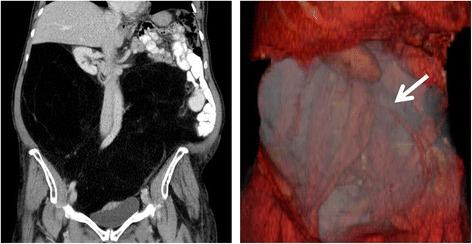
Figure 1.

The tumor on computerized tomography and on 3D-reconstruction. On the left the tumor is displayed on coronal plane, showing massive shifting of the intestines and kidneys. On the right the tumor is shown on 3D-reconstruction, nearly filling the whole abdomen with encasement of the inferior mesenteric artery (white arrow).
