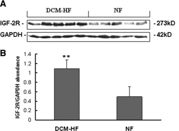
Fig 1.

Western blot analysis of cardiac IGF-2R. (A) Representative Western blot analysis of cardiac IGF-2R expression. (B) Densitometric quantification of cardiac IGF-2R expression. The IGF-2R protein levels related to the internal standard protein GAPDH were calculated as the relative abundance. Densitometric analyses of the blots showed an apparent increase in IGF-2R in DCM failing hearts (DCM-HF, n = 5) compared with non-failing control hearts (NF, n = 5; **P < 0.01). Data are presented as mean ± S.D.
