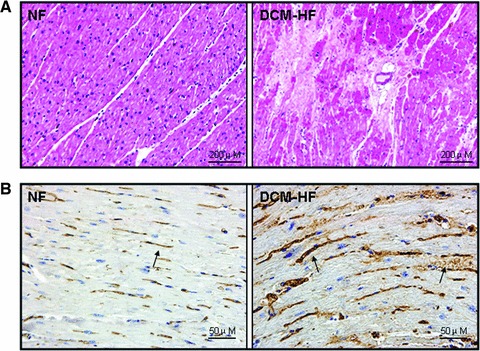
Fig 2.

Histopathological analysis of IGF-2R. (A) Representative light microscopic findings in haematoxylin and eosin staining. The normal appearance of myocardial fibres with central nuclei is seen in non-failing control hearts (NF) and interstitial fibrosis replacement is found in DCM failing hearts (DCM-HF). (B) Representative light microscopic IGF-2R immunoreactivity using a monoclonal antibody, which recognizes IGF-2R. Few and weak immunoreactivity of IGF-2R is observed in non-failing control hearts (NF), but more and strong IGF-2R immunoreactivity can be seen in DCM failing hearts (DCM-HF). Arrows indicated the positive staining.
