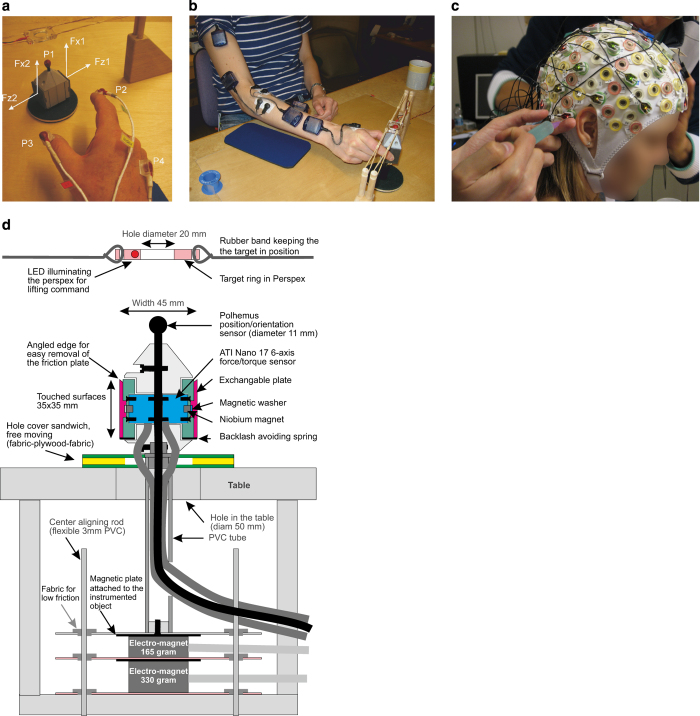
Figure 1. Methods.
(a) Force and position sensors. F1-F2 correspond to force/torque sensors (ATI Nano), with x corresponding to lift force and z to grip force. P1-P4 correspond to 3D position sensors (Polhemus FASTRAK) attached to the object (P1), the index finger (P2), the thumb (P3) and the wrist (P4). (b) EMG sensor placement: 1-anterior deltoid, 2-brachioradialis, 3-flexor digitorum, 4-common extensor digitorum, 5-first dorsal interosseus. (c) EEG sensor (ActiCap), recording from 32 electrodes. (d) Test object. The object to be grasped was visible on top of the table (cf, panel a) while the rest was hidden from view. The distance between the two contact surfaces (each 35×35 mm) measured 45 mm and they were secured to the object by niobium magnets. The touched surface could easily be replaced. The force applied to each contact plate was measured with mechanically isolated ATI Nano 17 6-axis force/torque sensors. The weight of the object including its magnetic plate was 165 g and could be increased to 330 or 660 g by controlling two electromagnets at the bottom. A set of flexible PVC rods provided low-friction alignment of the object on the table. Above the object was a rectangle in perspex with a centre hole (Ø20 mm). The start of the trial was signalled when the Perspex rectangle was illuminated by a LED. On top of the object was a Polhemus sensor mounted to record the position and orientation of the object.

