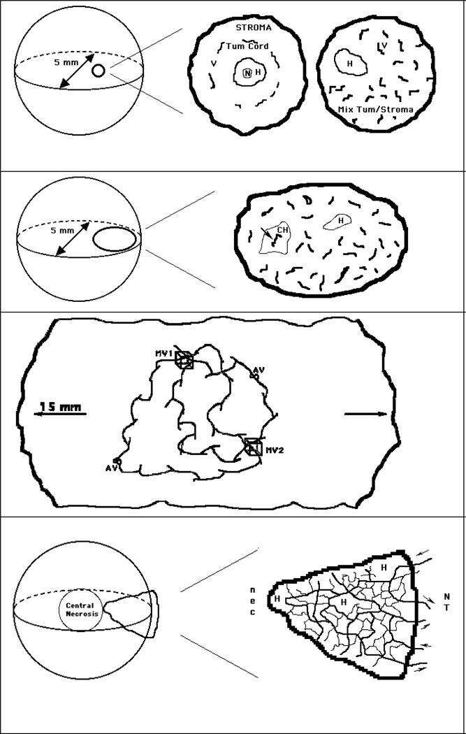
Figure 1.
a - Diffusion-Limited Hypoxia - This type of hypoxia (H) occurs when the spatial heterogeneity of vessels causes small zones of tumor tissue to be too far from vessels for diffusion of oxygen. It was initially described in epithelial tumors where cords of tumor tissue are surrounded by vessels within the normal stroma (center) but is also found in non-epithelial tumors (right). Sometimes, necrosis (N) is found in the center of a hypoxic zone. The centre and right panels are an expanded view of a 0.5 mm sphere from a 5 mm diameter tumor (left).
b - Cycling Hypoxia - This type of hypoxia (CH) occurs when blood flow in an isolated blood vessel cycles in an on-off pattern (arrow). It was proposed that two fluorescent DNA-binding dyes, when injected at different times, could label such vessels by demonstrating only one of the two dyes. A region of diffusion-limited hypoxia (H) is also shown. Right panel represents an expanded view of a 1.5 mm ovoid from a 5 mm diameter tumor, left).
c - In larger and particularly orthotopic tumors, blood travels over extended distances along unpaired vessels, which originate from co-opted normal-tissue vessels. This allows for a very heterogeneous mixture of vessels that can be carrying hypoxic or aerobic blood depending on the distance from the nearest arterial source. In this highly simplified diagram, two artery-vein pairs are depicted (AV) in a 15 mm diameter tumor. These produce various types of paths that may travel through volumes of interest (MV1 & MV2). In this example MV1 is perfused by predominantly arterial blood, whereas MV2 is perfused by predominantly venous blood. Note the 15 mm scale, vs 5mm for the other figures.
d - In smaller (5 mm) murine tumors (as described in panels a & b) vessels that arise from the normal tissue (NT) in the tumor periphery may be incapable of carrying oxygen and nutrients to the tumor interior. This can lead to macroscopic central necrosis (nec) - a feature often observed in such tumors. In this highly simplified diagram (expanded in right panel), a network of vessels is shown to originate from 2 arterioles and venules (arrows) outside a wedge-shaped portion of tumor. In an actual tissue section of such a region, many more ‘vessels’ would be observed since the network is not planar and the tissue section would cut randomly through these and other vessels/vessel-networks. Small regions of hypoxia (H) are also shown.

