
Figure 3.
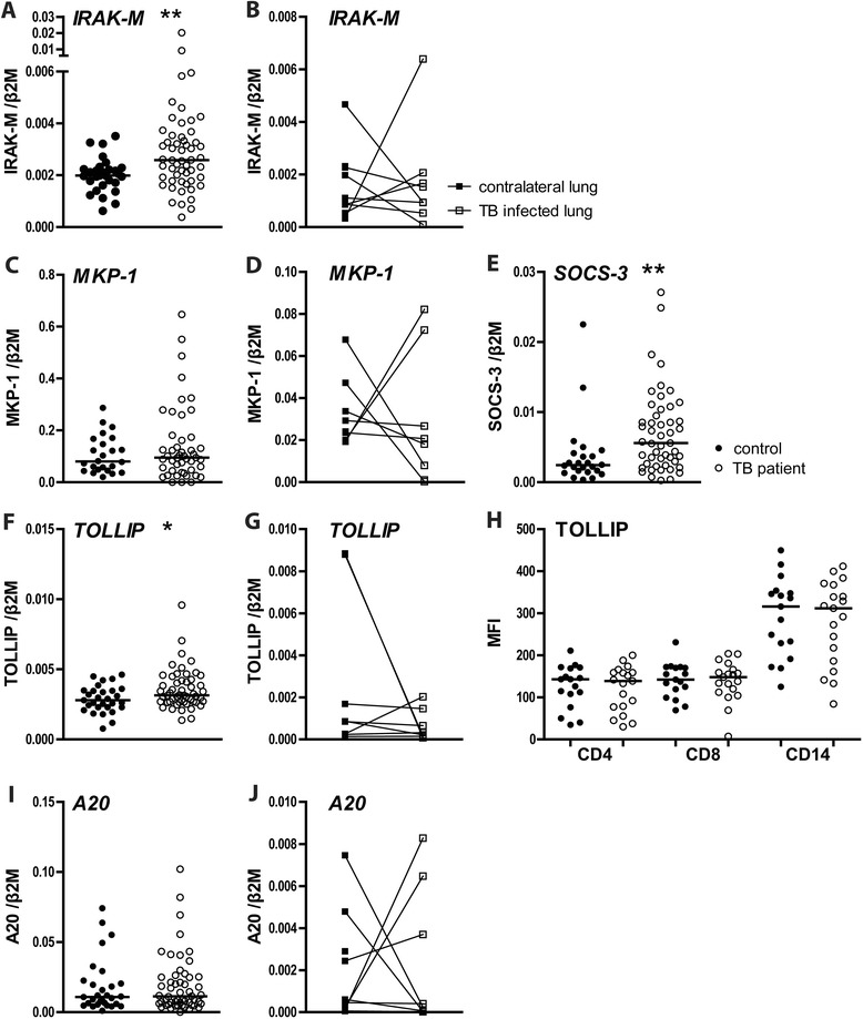
Intracellular negative regulators and mediators of TLR signaling. mRNA levels in PBMCs of TB patients (open dots) and healthy controls (closed dots) and TB patient AMs from the diseased (TB-positive) lung segment (open squares) and from the matching contralateral lung (closed squares). PBMC mRNA expression of IRAK-M (A), MKP-1 (C), SOCS-3 (E), TOLLIP (F) and A20 (I). AM mRNA expression of IRAK-M (B), MKP-1 (D), TOLLIP (G) and A20 (J). TOLLIP (H) expression levels in CD4 or CD8 positive lymphocytes and CD14 positive monocytes, as determined by flow cytometry (mean channel fluorescence intensity, MFI). mRNA expression is normalized to β2-microglobulin. Depicted are dot plots with medians; open dots: TB patients, closed dots: healthy donors. *P < 0.05, **P < 0.01.