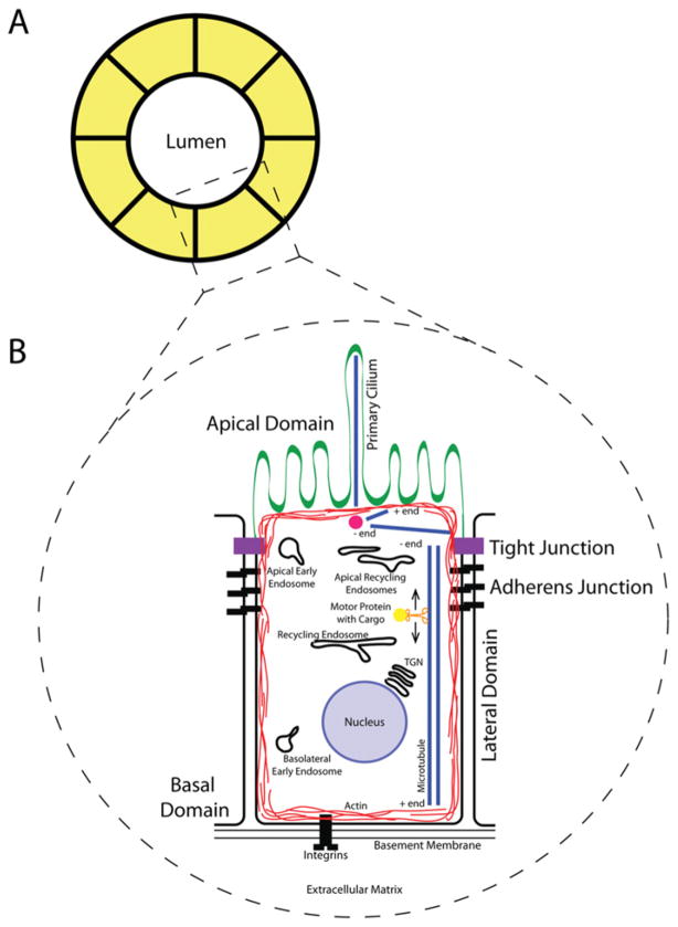
Figure 1. Structure of the mammalian epithelia.
(A) Cross-section of a polarized cyst or tubule. The apical domain of the PM faces the hollow lumen, and the basolateral domain faces the ECM. (B) Schematic representation of a single polarized epithelial cell. The apical domain faces the lumen and contains the specialized subdomain, the primary cilium. The tight junction separates the apical and basolateral domains and is composed primarily of occludins and claudins. Cadherins and nectins make up the adherens junction, which lies directly basal to the tight junction, and functions as a link to the actin cytoskeleton, which forms a cortex around the cell’s periphery. The lateral domain of the cell faces neighbouring cells in the monolayer, while the basal domain faces the basement membrane and interacts with the ECM via integrins. Microtubules are oriented with their plus end facing the apical domain and their minus end facing the basal domain. Motor proteins transport cargo via endocytic carriers along these microtubules.

