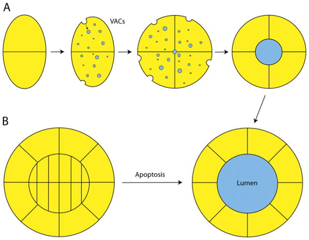
Figure 3. Models of epithelial lumen morphogenesis.
(A) In the hollowing model of lumen morphogenesis, newly polarizing cells divide, and, at the two-cell stage, the basolateral domain is the site of cell–cell contact, while the apical domain faces the ECM. Endocytosis of apical proteins and ECM fluids occurs through the use of specialized organelles called VACs, which are targeted to the meeting point of the dividing cells. These VACs accumulate and form the apical lumen at the centre of the forming cyst. Glycoproteins and polysaccharides are transported in these VACs and aid in the self-repulsion of the apical PM, allowing the lumen to remain open. (B) In the cavitation model, cells proliferate to form a solid cyst or tube. The outer cells of this structure, which are in contact with the ECM, then polarize. The cells internal to this monolayer then undergo apoptosis, resulting in the clearing of the apical lumen.

