Figure 1.
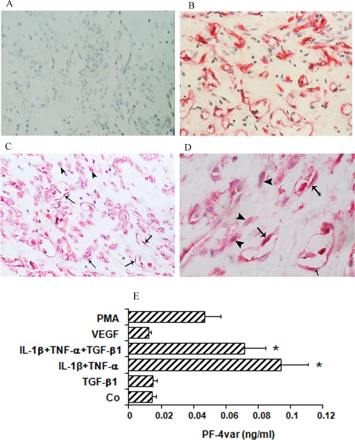
Expression of PF-4var/CXCL4L1 in proliferative diabetic retinopathy (PDR) and stimulated human retinal microvascular endothelial cells (HRMEC). Epiretinal membranes from patients with PDR were subjected to immunohistochemistry using antibodies against CD34 and PF-4var/CXCL4L1. (A) Control slide that was treated with an irrelevant isotype control antibody showing no labeling. (B) Immunohistochemical staining for CD34 showing blood vessels positive for CD34. (C, D) Immunohistochemical staining for PF-4var/CXCL4L1 showing immunoreactivity in vascular endothelial cells (arrows) and stromal cells (arrowheads). Low power (C) (original magnification ×40) and high power (D) (original magnification ×100). To verify whether HRMEC can produce PF-4var/CXCL4L1 in vitro as well, HRMEC were incubated for 96 hours with 100 ng/mL phorbol myristate acetate (PMA), 10 ng/mL VEGF, 10 ng/mL TGF-β1, 10 ng/mL IL-1β plus 30 ng/mL TNF-α, or 10 ng/mL IL-1β plus 30 ng/mL TNF-α plus 10 ng/mL TGF-β1 or were left untreated (Co) as described in Materials and Methods (E). Results represent the mean (± SEM) PF-4var/CXCL4L1 concentration measured by ELISA (*P < 0.05) (n = 5).

