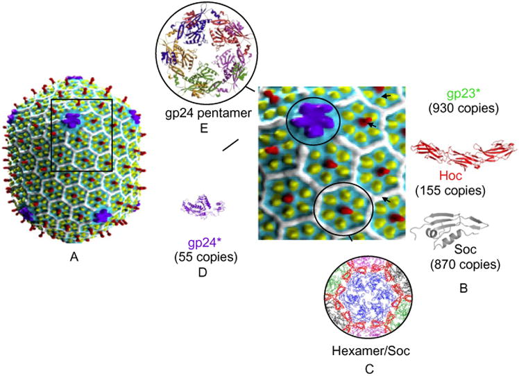
Figure 1.

Structure of the bacteriophage T4 head. (A) CryoEM reconstruction of phage T4 capsid (Fokine et al., 2004); the square block shows enlarged view showing gp23 (yellow subunits), gp24 (purple subunits), Hoc (red subunits), and Soc (white subunits). (B) Structures of RB49 Hoc (red) and RB69Soc (gray). (C) Structural model showing one gp23 hexamer (blue) surrounded by six Soc trimers (red). Neighboring gp23 hexamers are shown in green, black, and magenta (Qin et al., 2009). (D) Structure of gp24 (Fokine et al., 2005). (E) Structural model of gp24 pentameric vertex.
