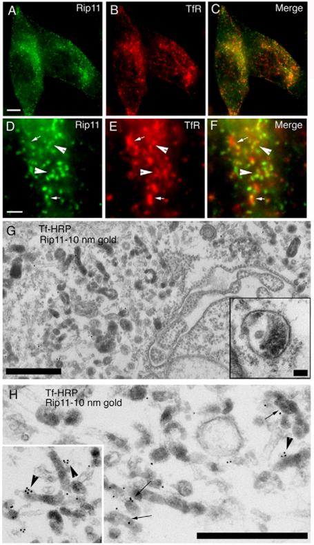
Fig. 1.
Rip11/FIP5 is enriched in peripheral recycling endosomes. (A-F) HeLa cells were plated on collagen-coated glass coverslips fixed and stained with anti-TfR (B,C,E,F, red) or anti-Rip11/FIP5 (A,C,D,F, green) antibodies. Yellow in C and F represents overlap between TfR and Rip11/FIP5. (D-F) High-magnification images of the same cell. Arrowheads in D-F indicate organelles where TfR and Rip11 colocalize. Arrows in D-F indicate organelles where Rip11/FIP5 is enriched in subdomains. Scale bars: 5 μm (A) and 1 μm (D). (G,H) HeLa cells were incubated with transferrin-HRP for 45 minutes at 37°C before being processed for immunoelectron microscopy. Anti-Rip11/FIP5 antibodies were detected using 10 nm gold. The electron-dense DAB reaction product indicates the presence of Tf. Clusters of Tf-positive endosomes that are also positive for Rip11/FIP5 are visible. Scale bars: 500 nm. Inset in H is a higher magnification image of endocytic tubules. Inset in G shows a vesicular part of an early endosome that is negative for anti-Rip11/FIP5 antibodies. Scale bar: 100 nm. Arrows indicate Tf-HRP-containing endosomes that are also positive for Rip11-gold. Arrowheads indicate Rip11 clusters on Tf-HRP endosomes.

