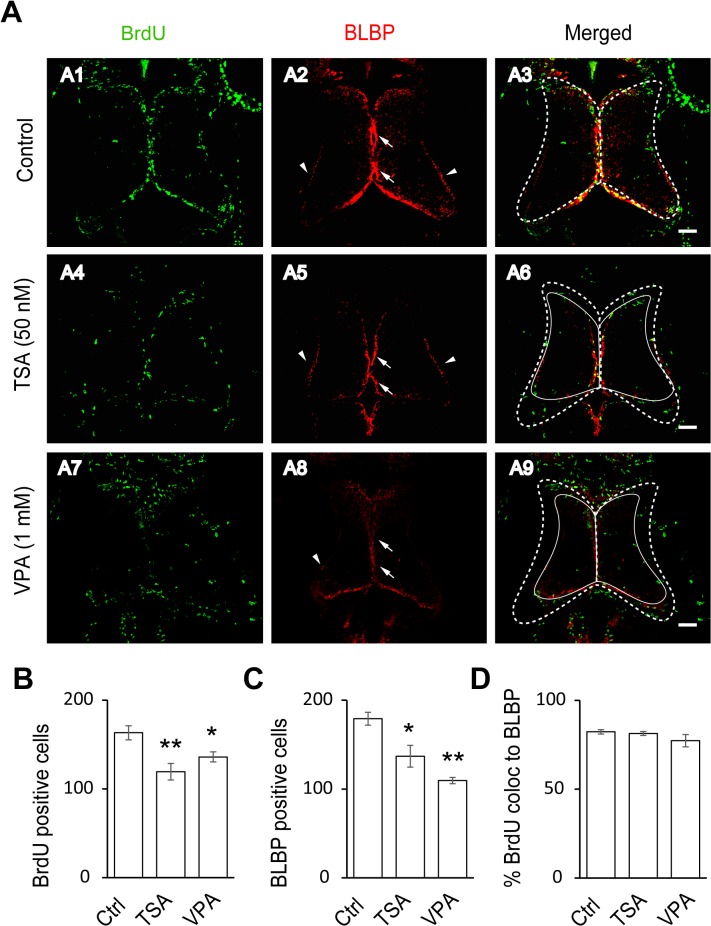
Fig 3. HDAC inhibitors block the proliferative rate of radial glia cells.
(A). Representative co-staining images showing the BrdU- and BLBP-positive cells in control (A1–A3), TSA-treated (50 nM, A4–A6) and VPA-treated (1 mM, A7–A9) tecta. The BLBP-positive cell bodies reside along the midline of the ventricular layer of the tectum (arrows) with endfeet on the edge of neuropil (arrow heads). The control tectum was outlined with a white dotted line (A3), which was put on TSA- (A6) or VPA-treated (A9) tectum. The TSA- (A6) or VPA-treated (A9) tectum was outlined with a solid line, which is smaller than control tectum (A3). Scale: 50 μm. (B-C) Quantification data showing that the number of BrdU- and BLBP-positive cells were significantly decreased in TSA- or VPA-treated tecta compared to the control. (BrdU: Ctrl, 163.2 ± 7.9, N = 5, TSA, 119.4 ± 9.3, N = 5, VPA, 136.0 ± 5.7, N = 3; BLBP: Ctrl, 179.2 ± 7.2, N = 5, TSA, 137.0 ± 12.2, N = 5, VPA, 109.7 ± 3.5, N = 3; *p<0.05, **p<0.01). (D). Most of BrdU-labeling cells are colocalized to BLBP-positive cells (Control: 82.3% ± 1.2%, N = 5, TSA: 81.4% ± 1.1%, N = 3, VPA: 77.3% ± 3.4%, N = 3).

