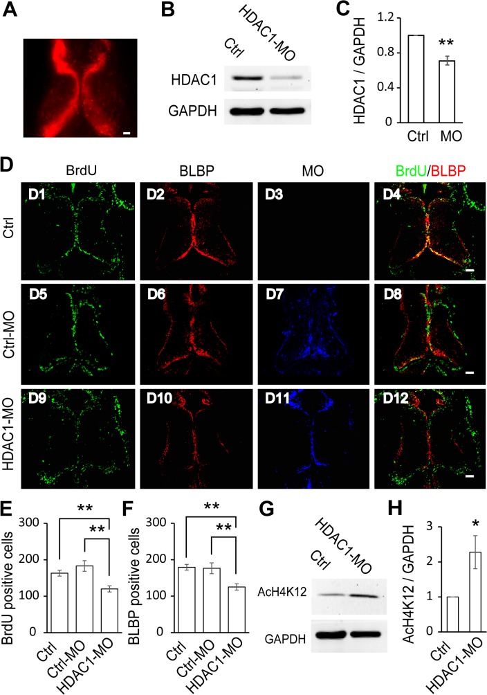
Fig 4. HDAC1 knockdown decreases cell proliferation in the optic tectum.
(A). Representative fluorescence image showing the optic tectum transfected with HDAC1-MO tagged with lissamine in vivo. (B). Western blot analysis of homogenates from control and HDAC1-MO transfected brains using an anti-HDAC1 antibody. (C). Quantification revealed that HDAC1 expression was significantly decreased in the HDAC1-MO transfected tectum compared to controls. Data is represented as an intensity ratio of HDAC1 to GAPDH normalized to the control value. Two-tailed T-test, N = 3, **p<0.01. (D). Representative immunofluorescence images of BrdU- and BLBP-labeled cells in control (D1-D4), Ctrl-MO (D5-D8), and HDAC1-MO (D9-D12) transfected brains in stage 48 tadpoles. Scale: 50 μm. (E-F). Summary data showing that HDAC1-MO transfection significantly decreased the number of BrdU- (E) and BLBP-labeled cells (F). There was no significant change in BrdU- or BLBP-labeled tectal cells electroporated with Ctrl-MO (E, F). (BrdU: Ctrl, 163.2 ± 7.9, N = 5, Ctrl-MO, 183.0 ± 14.6, N = 4, HDAC1-MO, 120.0 ± 8.5, N = 5; BLBP: Ctrl, 179.2 ± 7.2, N = 5, Ctrl-MO, 176.5 ± 11.5, N = 4, HDAC1-MO, 125.2 ± 8.4, N = 5; **p<0.01). (G). Acetylation levels of histone H4 at lysine 12 (AcH4K12) were measured by Western blot of total optic tectal extracts. Representative bands for control and HDAC1-MO transfected tadpoles. (H). Summary data showing that acetylation of H4K12 in HDAC1-MO animals is significantly increased compared to control tadpoles. N = 3, Two-tailed T-test, *p<0.05.

