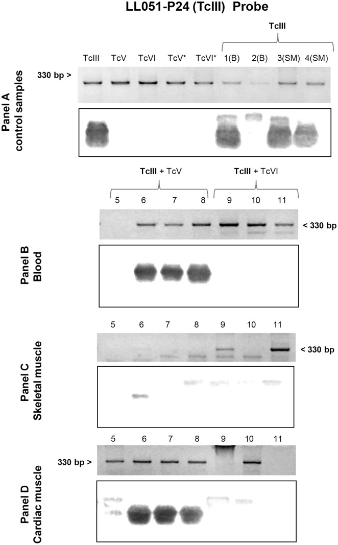
Fig 3. Southern blot analyses using the TcIII (LL051-P24) probe.
Each panel shows the electrophoretic pattern of minicircle regions and kDNA transferred to a nylon membrane. Panel A: lane TcIII, TcV, TcVI, TcV* and TcVI* correspond to DNA of parasite culture from LL051-P24 (DTU TcIII), LL055R3cl2 (DTU TcV), CL-Brener (DTU TcVI), LL014–1 (DTU TcV*) and LL040–1 (DTU TcVI*) respectively; and lane 1–4: blood (B) and skeletal muscle (SM) samples of mouse infected with TcIII isolate. The asterisk as superscript of the DTU indicates DNA sample from culture of the same inoculated isolate. Panel B, C and D: blood, skeletal muscle and cardiac muscle, respectively, of animals infected with TcIII + TcV (Lane 5–8) and TcIII + TcVI (Lane 9–11).

