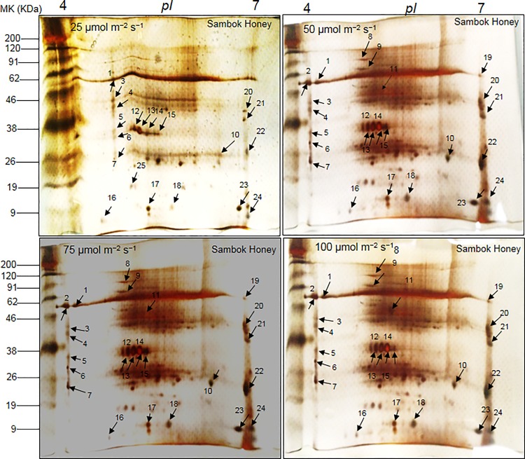
Fig 2. 2-DE reference maps of vascular connections (connected portions) in watermelon (Citrullus vulgaris Schrad.) ‘Sambok Honey’ as the scion and bottle gourd (Lagenaria siceraria Stanld.) ‘RS Dongjanggun’ as rootstock seedlings grown under different photon flux densities (25, 50, 75 and 100 μmol m−2 s−1 PPFD).
Proteins from vascular connections (70 μg) were elctrofocused on a pH 4–7 IPG strip (11 cm), separated onto 12.5% (w/v) SDS-PAGE. The gels were silver stained and visualized as described in experimental section. Protein spots marked by arrows were identified by MALDI-TOF/TOF-MS as described in experimental sections.

