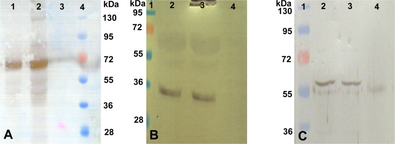
Fig 7. Western blot analysis of 5 days cultured buffalo hepatocytes.
Blots of buffalo hepatocytes lysate by using antibodies against albumin (Panel A); lane 1: HepG2 (Positive control), Lane 2: buffalo hepatocytes (test); Lane 3: skin fibroblast (negative control); Lane 4: pre-stained protein marker; cytokeratin-18 (Panel B); Lane 1: pre-stained protein marker; Lane 2: HepG2 cells; Lane 3: buffalo hepatocytes; Lane 4: skin fibroblast; and α1-antitrypsin (Panel C), order of lanes is similar to that shown in panel B.

