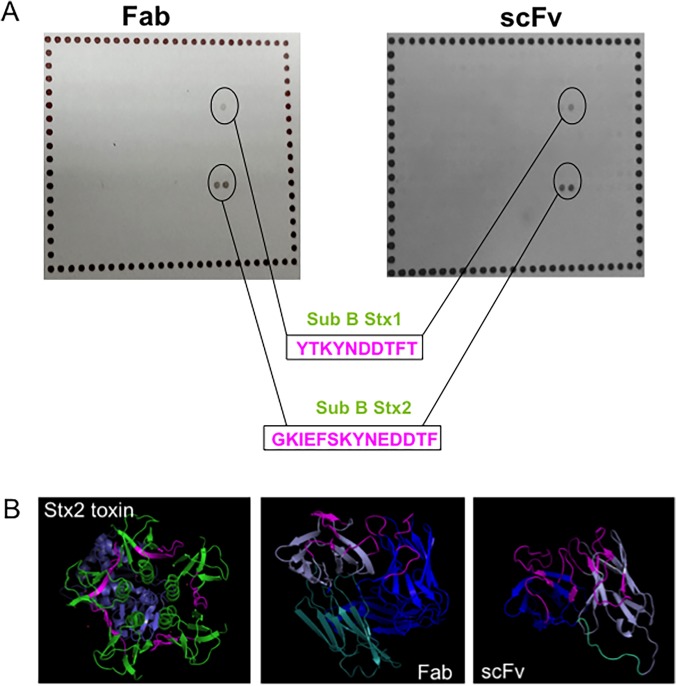
Fig 3. Structural analyses of antibody fragments.
A. Peptide mapping and the corresponding peptides, with the epitopes highlighted in pink. B. Structure prediction of both recombinant antibodies and Stx2, subunit A (purple) and subunit B (green), with the recognized peptides highlighted in pink as well as the antibody CDRs of recombinant antibodies. Also, the variable chains are represented, heavy (blue) and light (purple). In the Fab structure, the constant chain is shown in light blue, as well as the scFv linker.

