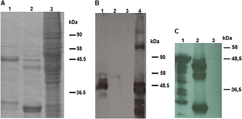
Fig 1. SDS-PAGE-Coomassie staining and Western Blot of HPV18L1–45RG1 VLP.
Purified and dialyzed 18L1–45RG1 VLP (lane 1), HPV18 wt L1 VLP (lane 2) or crude Sf9 lysate (lane 3) were separated by SDS-PAGE followed by Coomassie staining (A). The L1-RG1 fusion protein migrated at a molecular weight of about 50kDa, slightly slower than wt HPV18 L1, for which smaller degradation products are also visible. Insertion of the RG1 peptide into the L1 protein and its antigenicity were verified by Western Blot using an anti-HPV16 L2 aa 11–200 serum (B), or Camvir-1 reacting to HPV18 L1 (C). For both Western Blots, HPV18L1–45RG1 fusion proteins show a molecular weight of about 50kDa (Fig. 1B lane 1; Fig. 1C lane 1), with smaller bands representing proteolytic degradation products. As controls HPV18 wt L1 VLP (Fig. 1B lane 2; 1C lane 2), HPV16L1L2 VLP (Fig. 1B lane 4) and Sf9 cells only (Fig. 1B lane 3; Fig. 1C lane 3) were used.

