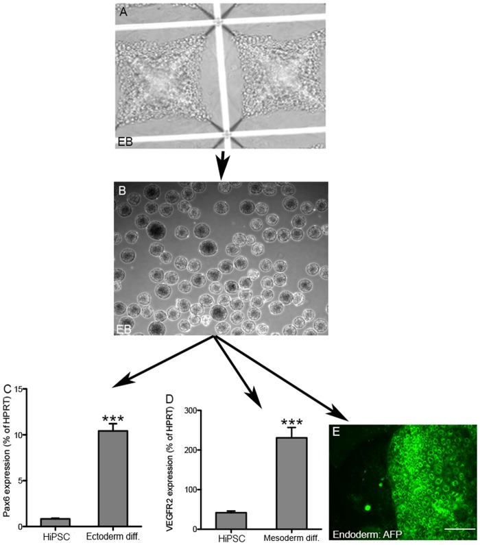
Fig 3. Differentiation of HiPSCs to embryoid bodies (EB) and germ layers.
The ability of HiPSCs to form EB was demonstrated by dispersing colonies to single cell suspensions and placing them in aggrewell plates (A). After 24 hours, homogenous EB were formed (B). EB were differentiated to three germ layers as described in the Materials and Methods. (C and D) Germ layer formation was demonstrated by RT-PCR (C, Pax6 for ectoderm, and D, VEGFR2 for mesoderm markers respectively). (E) The endoderm marker, alpha-fetoprotein (AFP), was detected by immunofluorescence. Scale bar: 200 μm. The experiments in panels C and D were repeated 3 times; P<0.001.

