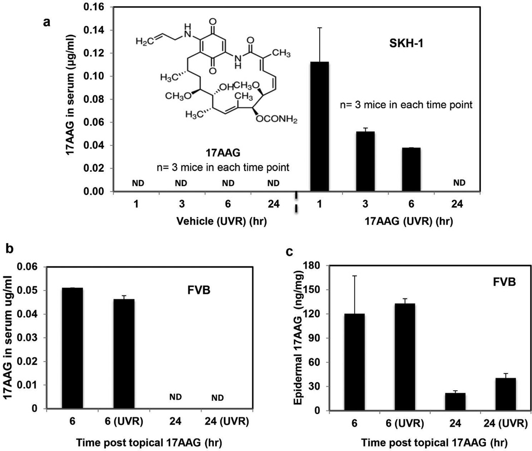
Figure 2. Topically applied 17AAG to skin is distributed both in epidermis and serum.
(a) The SKH-1 hairless mice (6–7 weeks old) were exposed to UVR (1.8 kJ/m2) three times weekly (Monday, Wednesday and Friday). The mice in the vehicle group (n=3) received topical treatment of 200 µl vehicle (DMSO:acetone: 1:40 v/v) before and after UVR exposures. The mice in 17AAG group (n=3) received freshly prepared 500 nmol of 17AAG (DMSO:acetone: 1:40 v/v) before and after each UVR exposure. All mice were treated for 25 weeks and sacrificed at 1, 3, 6 and 24 h post last UVR exposure. Blood samples were collected to analyze serum 17AAG by HPLC. (b, c) In a separate experiment, wild type FVB mice (6–7 weeks old) were treated once topically with either 17AAG alone (n=4) or in conjunction with a single UVR (1.8 kJ/m2) exposure (n=4). Mice of both the groups were sacrificed at 6 h (n=2) and 24 h (n=2) post 17AAG treatment. Blood samples were collected and epidermal protein lysates were prepared for 17AAG analysis. To prepare epidermal lysate, epidermis was scraped and homogenized in the lysis buffer. For 17AAG analysis, 50µl of epidermal lysate was used. Bar graph illustrates the serum (a,b) and the epidermal (c) 17AAG levels. The epidermal 17AAG values were normalized with total protein concentration.. Values shown in all bar graphs are mean±SE. ND: Not detectable. Inset: Chemical structure of 17AAG.

