
Figure 4.
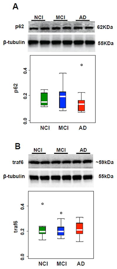
(A, B) Representative immunoblots and box plots of hippocampal levels of p62 (A) and traf6 (B) in cases clinically diagnosed as NCI, MCI and AD. β-tubulin was used to normalize the immunoreactive signals obtained in the blots by densitometry. Levels of p62 and traf6 were stable across clinical groups. NCI, non-cognitive impairment, MCI, mild cognitive impairment, AD, Alzheimer disease. Circles in the box plots indicate outliers.