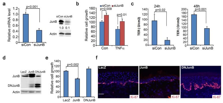
Figure 1. JunB loss-of-function leads to increased epidermal cell proliferation and reduced barrier function.
(a) Confirmation of gene silencing via real-time RT-PCR and Western blotting. Relative expression levels of JunB were shown below each band via densitometry. (b) Cell growth of human keratinocytes treated with or without 50ng/ml TNFα for 72h. (c) Barrier functions as measured by the Transepithelial Electrical Resistance (TER). Keratinocytes were induced to differentiate with 1.2 mM Ca++on permeable membranes for 24h and 48h prior to TER measurement. (d) Confirmation of gene expression via immunoblotting. (e) Cell growth analysis. (f) Immunostaining of 6-week old skin grafts generated on SCID mice with human keratinocytes transduced to express genes as indicated. Ki-67 [orange] and nuclei [blue]. Graphs represent the average values of triplicate samples + SD. P-values were obtained via two-tiered student T-test. Scale bar=100μm.

