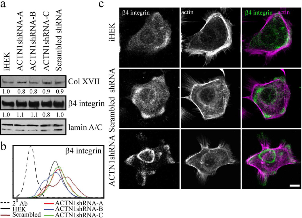
Figure 3. ACTN1 knockdown and effects on hemidesmosomal protein expression and localization.
(a) Extracts of iHEKs, the three ACTN1 knockdown clones (ACTN1shRNA-A, -B and -C) and iHEKs expressing scrambled shRNA were processed for immunoblotting using antibodies against collagen XVII (Col XVII), β4 integrin or lamin A/C as indicated. Blots were scanned and quantified by densitometry, values were normalized to lamin A/C levels and are displayed relative to iHEK levels. Lamin A/C reactivity was used as a loading control. The blot is representative of at least three independent trials. (b) The same cells as in a were prepared for FACS using antibodies against β4 integrin. 20 Ab indicates a control assay where primary antibody was omitted. (c) iHEKs, iHEKs expressing scrambled shRNA and iHEKs expressing ACTN1 shRNA were prepared for immunofluorescence staining with antibodies against β4 integrin together with rhodamine phalloidin. Panels on right show overlays of the two images. Bar, 10 µm.

