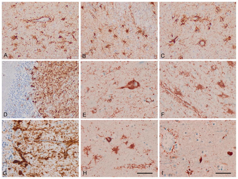
Figure 1.
Astrocyte-predominant familial tauopathy (case 1). (A) cerebral cortex; (B) striatum; (C) amygdala; (D) cerebellum; (E) neurons in the upper cerebral cortex; (F) astrocytes in the inner cerebral cortex; (G) cerebellum. (A–G) AT8; (H) 4Rtau; (I) 3Rtau. Note the morphology of reactive astrocytes and the presence of tau immunoreactivity in perivascular astrocyte foot processes surrounding blood vessels. Paraffin sections, with DAB chromogen (brown deposits) and lightly counterstained with hematoxylin. A–D, I, bar in I = 50 μm; E–H, bar in H = 25 μm.

