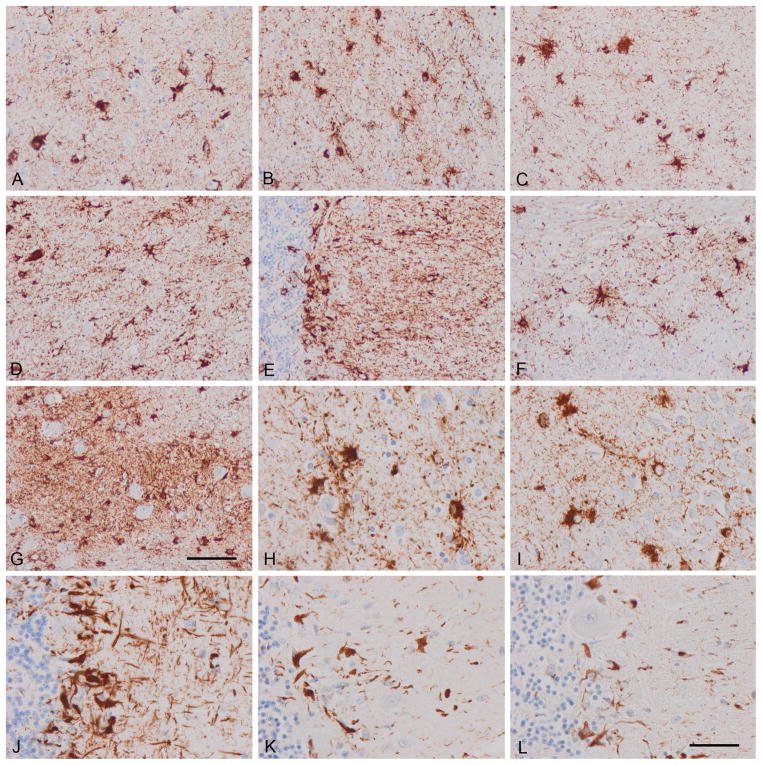
Figure 2.
Astrocyte-predominant familial tauopathy (case 2). (A) Upper layers cerebral cortex; (B) inner layers cerebral cortex; (C) CA1; (D) thalamus; (E) cerebellum; (F) pons; (G) inferior olive; (H) cerebral cortex; (I) dentate gyrus; (J–L) cerebellum. (A–J) AT8; (K) 4Rtau; (L) 3Rtau. Similar features as seen in case 1 showing the morphology tau-immunoreactive reactive astrocytes and perivascular immunoreactivity due to phosphor-tau deposition in astrocyte foot processes. Bergmann glia are heavily immunostained with anti-phospho-tau antibodies. Note the relatively small number of tau-containing neurons vs. the large number of tau-containing astrocytes. Paraffin sections, with DAB chromogen (brown deposits), lightly counterstained with hematoxylin; A–G, bar in G = 50 μm; H–L, bar in L = 25 μm.

