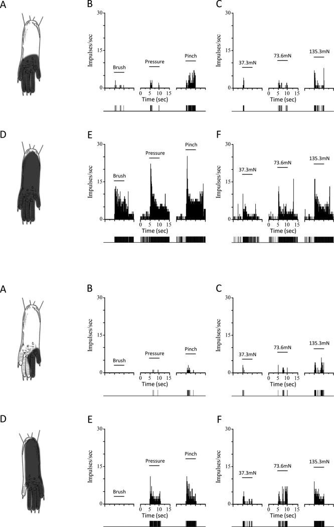
Figure 2.
Receptive field (RF) areas and responses of nociceptive neurons evoked by mechanical stimuli are greater in sickle mice. Upper panels: Location of the RF, and peristimulus time histograms showing discharge rates evoked by stimuli used for functional characterization (brush, pressure pinch applied to the RF) and responses evoked by von Frey monofilaments of controlled force for a single WDR neuron from a control (top panels A, B, and C) and from a sickle (lower panels D, E, and F) mouse. Solid horizontal lines represent the time of application of the stimuli (5 s). Discriminated output pulses from a window discriminator are provided below the histograms. Lower panels: Same format as above but RF areas and evoked responses are shown for single HT neurons from a control (A-C) and sickle (D-F) mouse. Bin width for all peristimulus time histograms is 100 ms.

