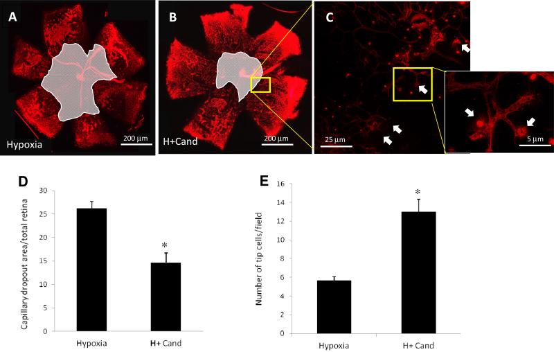
Figure 2. Candesartan stimulated number of tip-cells and enhanced reparative angiogenesis.
(A, B) Representative images of flat-mounted retinas of p17 animals subjected to hypoxia or hypoxia treated with candesartan (10mg/kg/day, ip) and labeled with isolectin-B4 (red). (C) Higher confocal magnified image (63X) of retinas labeled with isolectin B4 of the part corresponding to image B showing tip cells that grow toward the center of the retina to guide newly formed retinal capillaries. (D) Quantitative data of the capillary drop out area that were normalized to the total retina area showing that candesartan treatment reduced capillary dropout area by 45% in compared to that of untreated mice. (E) Quantitative data of the number of sprouting endothelial cell forming tip cells show that treatment with candesartan (10mg/kg) significantly increased the number of tip cellsare significantly higher among those treated with candesartan compared to untreated hypoxia. *p<0.05 vs hypoxia. (n=10-12/group).

