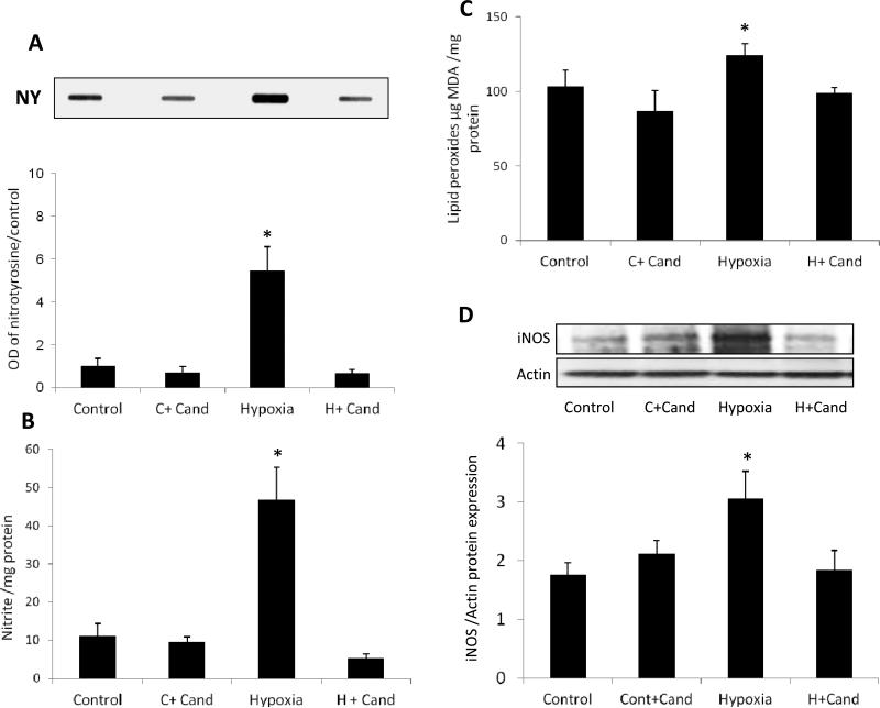
Figure 3. Candesartan blocked hypoxia induced oxidative and nitrative stress via suppression of iNOS.
(A) Representative image of slot blot and quantification for nitrotyrosine, the foot print of peroxynitrite. Hypoxia significantly induced nitrotyrosine formation compared to the normoxic control group. Candesartan treatment (10mg/kg/day,ip) significantly reduced the nitrotyrosine compared to untreated hypoxia (B) Nitrate level detected by Griess reagent which is an indirect measure of nitric oxide production in the retina, shows a significant increase in nitrate in hypoxia compared to the control groups which was blocked by candesartan treatment. (C) Lipid peroxidation detected by TBARs shows a significant increase in oxidative stress in hypoxia compared to the control groups, which was blocked by candesartan treatment. (D) Representative image and statistical analysis of western blot for iNOS normalized to β-actin showing thatiNOS expression is significantly increased in hypoxia compared to normoxia and normoxia-candesartan groups. Treatment with candesartan (10mg/kg/day,ip) reduced iNOS significantly compared to untreated pups. OD-optical density; NY –nitrotyrosine. *p<0.05 vs controls, n=4-6/group.

