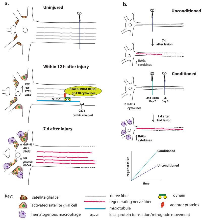
Figure 5.
The cell body response and the CL response. (a) The cell body response describes the morphological and molecular changes that occur in neurons after axotomy. Two types of signals reach the cell body from the site of injury. First, there is a very rapid Ca2+ wave, and, later, there are signals that have been retrogradely transported along microtubules via dynein and adaptor proteins. These different signals lead to changes in RAG expression in the axotomized cell body. In addition (though not shown), the accumulation of macrophages and the activation of satellite glial cells near the cell bodies leads to the release of extrinsic signals that also alter neuronal gene expression, for example, the secretion of gp130 cytokines by satellite glial cells in the SCG triggers an increase in galanin expression (Sun et al., 1994; Habecker et al., 2009; Zigmond, 2012b). Finally, in addition to the cell body response there is an alteration of protein translation in the axons themselves. (b) When a CL is made prior and also distal to a test lesion and neurite outgrowth is measured from the test lesion either in vivo or in vitro, growth is accelerated compared to when only a test lesion is made. As discussed in the text, macrophage accumulation in the region of axotomized cell bodies is required for a normal CL response.

