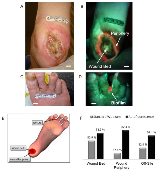
Fig 4. Autofluorescence detection of clinically-significant bacterial load in wound periphery and off-site areas.
A. White light image of a type II diabetic foot ulcer in a 78 y old female. B. Corresponding AF image showing heavy growth of S. aureus in the wound periphery missed by white light imaging. C. White light shows unremarkable areas between toes, while in D. the corresponding AF imaging detected bacterial biofilm, confirmed by microbiology. E. Schematic illustrates different wound locations where fluorescence imaging detected clinically significant bacterial load. F. Comparison of accuracy for correctly detecting clinically-significant bioburden between standard WL and AF imaging in wound bed, wound periphery and off-site areas. Scale bars: A,B. 1 cm; C. 2 cm, D. 1 cm.

