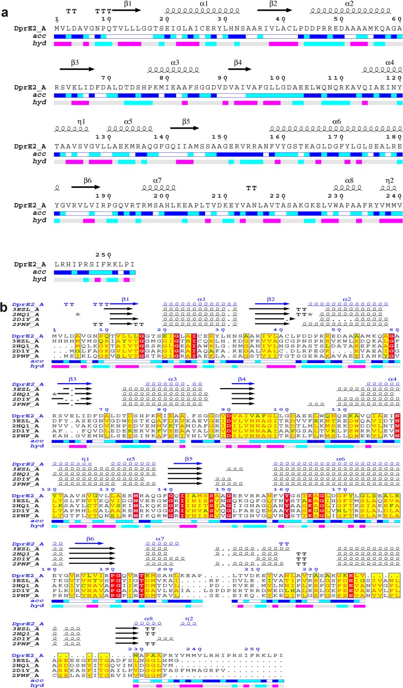
Fig 2. Secondary structure analysis and multiple sequence alignment between DprE2 and other members of the SDR protein family.
(a) Secondary structure analysis. The α helices, β sheets and turns are represented with squiggles, arrows and TT letters, respectively.The DprE2 accessibility is shown by color codes: blue for accessible, cyan is intermediate, white is buried. The hydropathy bar is shown by a bar where pink is hydrophobic and cyan is hydrophilic. (b) The multiple sequence alignment of DprE2 with its homologous SDR proteins is shown where identical and similar residues are boxed in red and yellow, respectively.

