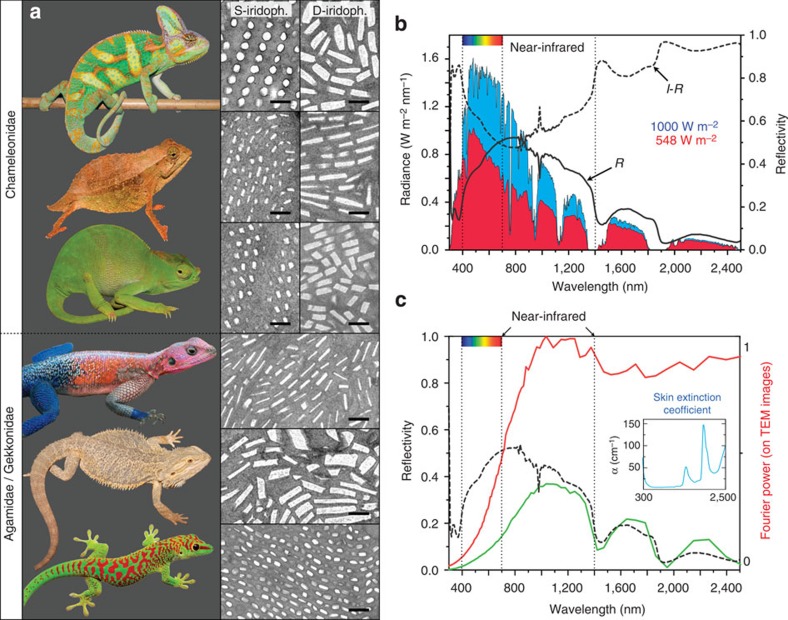
Figure 3. Iridophore types in lizards and function of D-iridophores in chameleons.
(a) In addition to F. pardalis (Fig. 1), other chameleonidae (top to bottom: Chamaeleo calyptratus, Rhampholeon spectrum and Kinyongia matschiei) exhibit two superposed layers of (S- and D-) iridophores, whereas agamids (the sister group to chameleons) and gekkonids have a single-type iridophore layer (top to bottom: Agama mwanzae, Pogona vitticeps and Phelsuma grandis). Scale bar, 500 nm. (b) Reflectivity (R) of a panther chameleon white skin sample and solar radiation spectrum (blue curve) at sea level (1,000 W m−2); the product of the solar radiation spectrum and (1−R) yields the amount of sun radiation absorbed by deep tissues (red curve, 548 W m−2). (c) The product of the Fourier power spectrum (red curve, computed from TEM images of D-iridophores) and the extinction coefficient of skin29 (blue inset) yields a predicted reflectivity distribution (green curve) similar to the measured reflectivity spectrum (black dashed curve) of a panther chameleon red skin sample.

