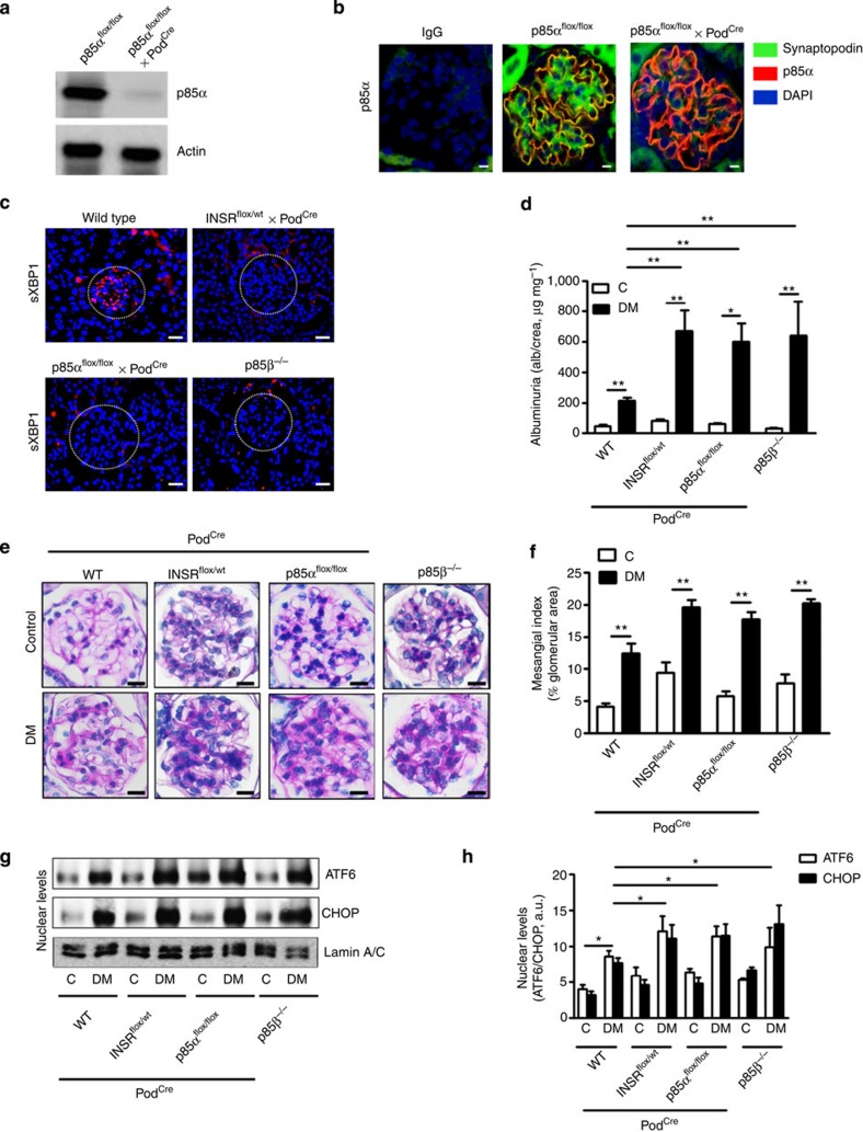
Figure 8. Signalling via INSR–p85 in podocytes is required for the adaptive ER-stress response in DN.
(a) Representative image showing immunoblot analysis of podocyte-specific nearly complete deletion of XBP1 and p85α in podocytes isolated from p85αflox/flox x PodCre mice when compared with podocytes isolated from control mice (p85αflox/flox mice). (b) Exemplary images showing podocyte-specific depletion of p85α in renal glomeruli of wild-type (p85αflox/flox) mice and p85αflox/flox mice crossed with PodCre. (c) Representative images showing sXBP1 levels (using an antibody detecting the spliced form of XBP1) in the renal glomeruli of non-diabetic wild-type mice and mice with podocyte-specific deletion of insulin receptor (INSRflox/Wt x PodCre) or p85α (p85αflox/flox x PodCre) or p85β deficiency (p85β−/−). (d–h) Bar graph summarizing albuminuria (d), representative images of the extracellular matrix deposition (e) bar graph reflecting extracellular matrix deposition (f), representative immunoblots (g), and bar graph (h) showing nuclear levels of ER transcription factors in renal cortex samples; (n=8 mice per group were analysed). C=control mice without diabetes, open bars; DM=diabetes, black bars. Mean±s.e.m. (d,f,h), *P<0.05, **P<0.01 (ANOVA).

