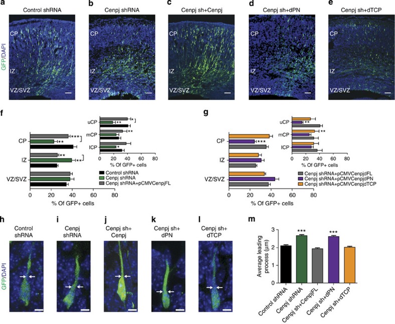
Figure 5. Microtubule-destabilizing domain of Cenpj is required for migration.
(a–e) Analysis of radial migration in cortices 3 days after co-electroporation of GFP with a control shRNA (a), Cenpj shRNA alone (b), Cenpj shRNA together with a full-length Cenpj expression construct (pCMV-Cenpj, c), Cenpj shRNA with a truncated Cenpj construct lacking the microtubule-destabilizing domain PN2-3 (pCMV-Cenpj dPN2-3, d) or Cenpj shRNA with a truncated Cenpj construct lacking the domain TCP (pCMV-Cenpj dTCP, e). Scale bar, 50 μm. (f,g) Quantification of the migration defects of GFP+ cells in the different zones of the cortex. The migration defect of Cenpj-depleted cells is rescued by overexpression of full-length Cenpj (f) and Cenpj dTCP but not of Cenpj dPN2-3 (g). Student’s t-test *P<0.05; **P<0.01; ***P<0.001. (h–l) Analysis of leading process thickness in neurons in the CP co-electroporated with GFP and Control shRNA (h), with Cenpj shRNA (i) and Cenpj shRNA and Cenpj full length (j) or Cenpj shRNA and Cenpj dPN (K) or Cenpj shRNA and Cenpj dTCP (l). Scale bar, 5 μm. (m) Quantification of leading process thickness in GFP+ neurons. The leading process enlargement observed in Cenpj-depleted neurons is rescued by co-expression of Cenpj shRNA with Cenpj full length and Cenpj dTCP but not by Cenpj dPN. Three embryos analysed for each condition; control shRNA, n=177 cells; Cenpj shRNA, n=200 cells; Cenpjsh+Cenpj FL, n=157 cells; Cenpj shRNA +Cenpj dPN2-3, n=274 cells; Cenpj shRNA+Cenpj dTCP, n=211 cells. Student’s t-test ***P<0.001. DAPI, 4',6-diamidino-2-phenylindole.

