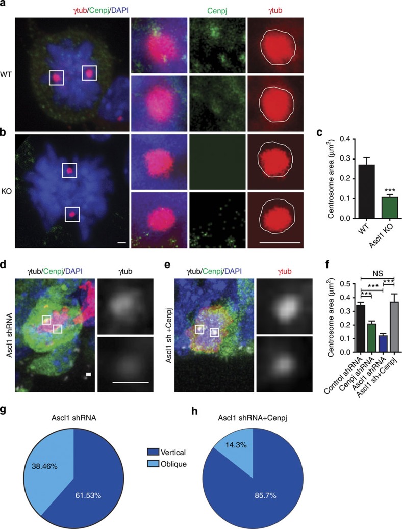
Figure 6. Cenpj is the main effector of Ascl1for centrosome biogenesis.
(a,b) Labelling of centrosomes in apical cortical progenitors from wild-type (WT) (a) or Ascl1 null embryos (b) with an antibody to γ-tubulin. Insets to the right show higher magnification of the boxed areas with separate colour channels. Scale bars, 1 μm. (c) Quantification of the area of the centrosomes in WT and Ascl1 null embryos labelled with an antibody to γ-tubulin. Three embryos analysed for each condition. Student’s t-test ***P<0.001. (d,e) Analysis of the centrosome area of GFP+-dividing apical progenitors electroporated 1 day earlier in utero with Ascl1 shRNA (d) or an Ascl1 shRNA plus a pCMV-Cenpj construct (e) labelled for GFP (green), pH3 (red) and γ-tubulin (grey). Insets to the right show higher magnification of the boxed areas with γ-tubulin (grey) channels. Scale bars, 1 μm. (f) Quantification of the area of the centrosomes in control shRNA-electroporated progenitors, Cenpj shRNA, Ascl1 shRNA and Ascl1 shRNA+pCMV-Cenpj-electroporated progenitors, labelled with γ-tubulin. The centrosomes are smaller in Ascl1-silenced progenitors and this defect is rescued in Ascl1-silenced cells co-electroporated with a Cenpj expression vector. Student’s t-test ***P<0.001. (g,h) Percentage of cells electroporated with Ascl1 shRNA (g) and Ascl1 shRNA+pCMV-Cenpj (h) having a vertical (60–90°) or oblique (30–60°) cleavage angle. Ascl1 shRNA, n=33 cells; Ascl1 shRNA+Cenpj, n=24 cells. Cells were analysed from at least six embryos obtained from three litters. DAPI, 4',6-diamidino-2-phenylindole; NS, not significant.

