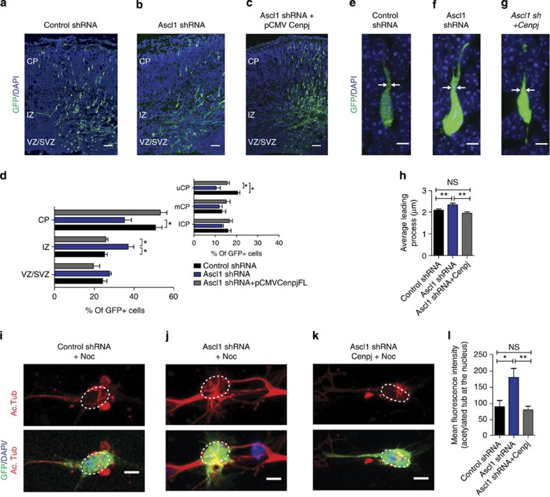
Figure 7. Cenpj is the main effector of Ascl1for microtubule regulation.
(a–c) Analysis of radial migration 3 days after electroporation of E14.5 cortices with GFP and a control shRNA, Ascl1 shRNA or Ascl1 shRNA+pCMV-Cenpj. Scale bar, 50 μm. (d) Quantification of the migration of GFP+ cells in the different zones of the cortex. The accumulation of Ascl1-silenced cells in the IZ but not their depletion from the uCP was rescued by overexpression of Cenpj. Student’s t-test *P<0.05 (e–g). Abnormal leading-process enlargement of Ascl1-silenced neurons in the CP. Scale bar, 5 μm. (h) Quantification of leading process thickness. Three embryos analysed for each condition; control shRNA, n=177 cells; Ascl1 shRNA, n=128 cells; Ascl1 shRNA+pCMV-Cenpj, n=239 cells. Student’s t-test **P<0.01. (i–k) Nocodazole treatment of cultured neurons 3 days after electroporation. Ascl1 silencing preserved a higher level of acetylated tubulin labelling in the cell body and leading process than control shRNA, and overexpression of Cenpj restored the acetylated tubulin-labelling intensity. Scale bar, 5 μm. (l) Quantification of acetylated tubulin labelling co-localized with the nucleus. Three independent cultures analysed for each condition; control shRNA, n=40 cells; Ascl1 shRNA, n=53; Ascl1 shRNA+pCMV-Cenpj, n=37. Student’s t-test *P<0.05; **P<0.01. DAPI, 4',6-diamidino-2-phenylindole; NS, not significant.

