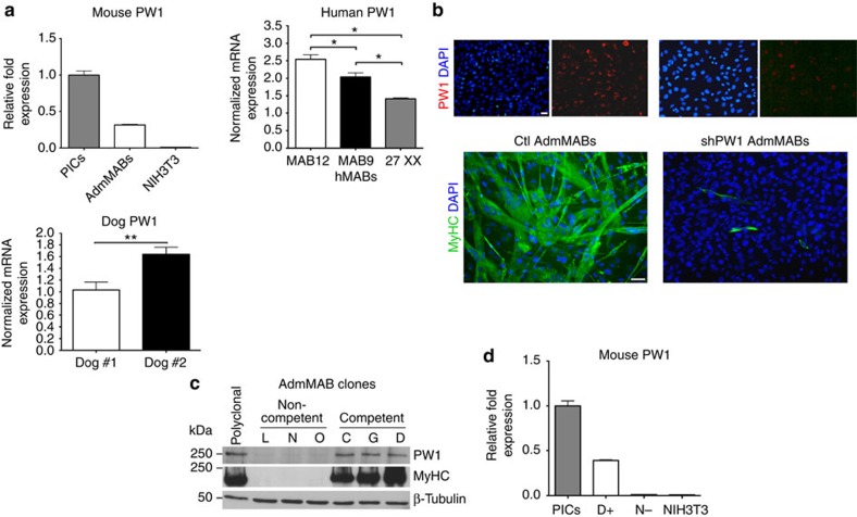
Figure 1. Silencing of PW1 interferes with mesoangioblasts (MABs) muscle differentiation.
(a) PW1 expression by qRT–PCR on different populations of mouse adult (AdmMABs), human and canine MABs. Values are plotted as relative messenger RNA (mRNA) expression and normalized to GAPDH levels. For the AdmMABs, values are expressed as fold expression relative to subpopulation of interstitial cells (PICs; =1). Each assay was performed in triplicate. Data are represented as means±s.d. *P<0.05, ns, not significant one-way unpaired t Test. (b) Immunofluorescence analysis for PW1 (red) and for the expression of all sarcomeric myosins (MyHC, green) on Ctl and shPW1 AdmMAB growing cells upon 5 days in differentiation medium. DAPI was used to stain nuclei. Scale bar represents 100 and 50 μm. (c) Western blot analysis of MyHC and PW1 expression on six different clones of AdmMABs isolated and selected for the different myogenic potency. Clones have been divided in competent (C, G, D) and non-competent (L, N, O) on the basis of their myogenic property. β-Tubulin was used to normalize the amount of loaded proteins. Polyclonal AdmMABs were used as a positive control. (d) PW1 expression by qRT–PCR on representative clones of competent (D+) and non-competent AdmMABs (N−). Values are normalized to GAPDH levels and expressed as fold expression relative to PICs (=1). NIH3T3 fibroblasts were used as negative control.

