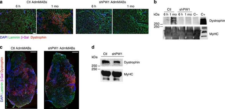
Figure 4. PW1-silenced MABs do not rescue the dystrophic phenotype after intra-arterial transplantation in scid-mdx mouse.
(a) Immunofluorescence staining for laminin (green), dystrophin (red), β-Gal (pink) and nuclei (DAPI, blue) on serial transverse sections of gastrocnemius muscle, 6 h and 1 month after intra-femoral artery injection of n-LacZ Ctl and shPW1 adult murine MABs (AdmMABs) into scid-mdx mice. Scale bar, 100 μm. n=4 for each group. (b) Western blot analysis of dystrophin expression in transplanted scid-mdx muscles, 6 h and 1 month (1 mo) after cell transplantation. C+ is a wt muscle, used as a positive control for Dystrophin expression; C− is an mdx-not transplanted muscle, representing the negative control for dystrophin expression. The expression of all the sarcomeric myosins, MyHC, was used to normalize the amount of loaded proteins. The C+ was loaded 10 times less to avoid Ab titration and photo bleaching. (c) Immunofluorescence staining for laminin (green), dystrophin (red), β-Gal (pink) and nuclei (DAPI, blue) on the transplanted tibialis anterior muscle, 1 month after intra-muscular injection of n-LacZ Ctl and shPW1 adult murine MABs (AdmMABs) into scid-mdx mice. n=4 for each group. Scale bar, 500 μm. (d) Western blot analysis of dystrophin expression in transplanted scid-mdx muscles, 1 month after cell transplantation. The expression of all the sarcomeric myosins, MyHC, was used to normalize the amount of loaded proteins.

