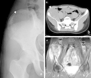
Fig. 3.

A 16-year-old patient with ES of the ilium. a A radiolucent lesion was visible in the right ilium on a plain X-ray (arrowhead). b CT detected a large mass that exhibited a periosteal reaction in the right ilium (arrowhead) and c, T2-weighted MRI depicted a mass that displayed heterogeneous high signal intensity in and around the right ilium and acetabulum
