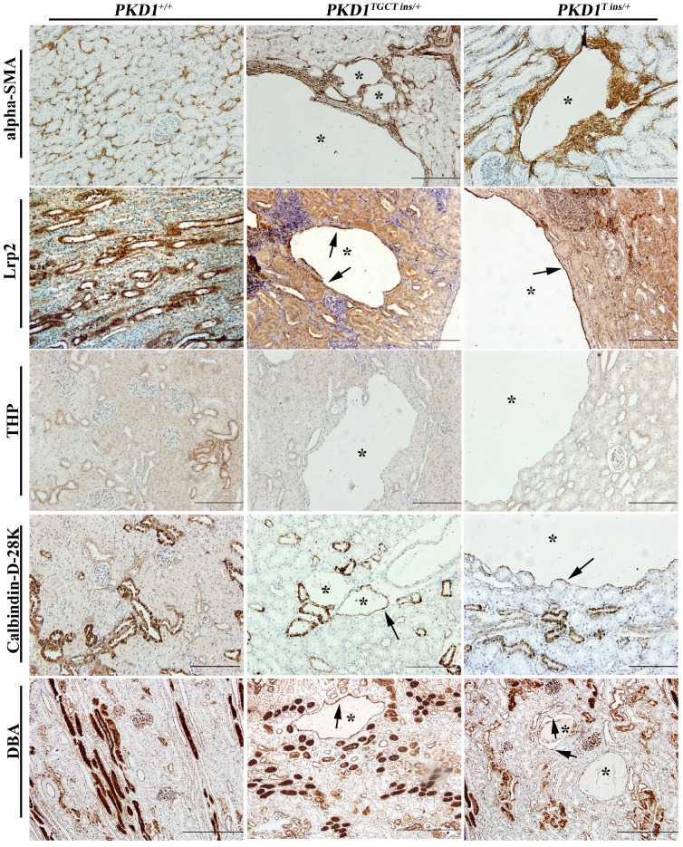
Figure 4.
Immunohistochemical characterization of renal cysts in PKD1+/- pigs. The upper panel shows that α-SMA staining fibrosis surrounding renal cysts (*) in the PKD1+/- pigs. The origin of renal cysts was determined using segment-specific markers/antibodies for proximal tubules (Lrp2), thick ascending limbs of loop of Henle/distal convoluted tubules (Tamm-horsefall protein, THP), distal convoluted tubules/cortical collecting ducts (Calbindin-D-28K), and collecting ducts (Dolichos Biflorus agglutinin, DBA). Cysts were positively stained by all of these antibodies/markers (arrow) except for THP. Each tubular segment specific marker was used to stain at least 30 cysts in each PKD1+/- kidneys. Scale bars in the images are 200 μm, except for DBA staining, which are 500 μm.

