
Fig. 3. Case 2, half lying-cone dog ear.
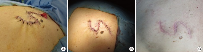
Sequenced images of the three-bite technique on the half lying-cone dog ear. (A) Reconstruction with a local skin flap and evidence of the dog ear at the end of the suture line. (B) The suture right after the three-bite technique. (C) The result at the 21-weeks follow-up.