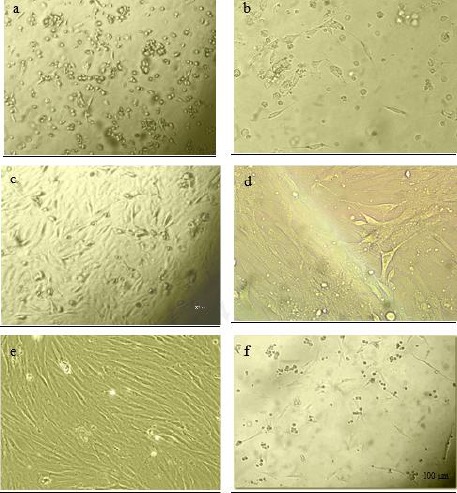
Figure 1.

Cell morphology of cultured rat derived omentum tissue mesenchymal stem cells were observed with an Olympus phase contrast microscope. The initial adherent cells appeared as separate colonies after 19 hr (a), and 2 days (b). More confluent omental stromal cells after 5 days (c) and monolayer cultured mesenchymal stem cells after one week (d). Spindle-shaped morphology of cells after new consecutive passages (e), and re-attachment of cells to the culture flasks following thawing after freezing stage (f)
