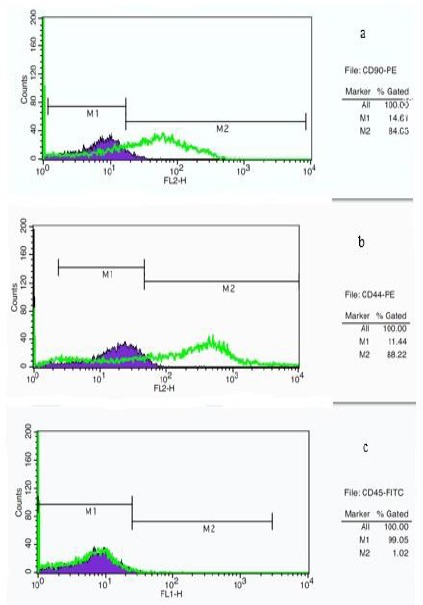
Figure 3.

Flow cytometry analysis of cell surface markers on rat derived omentum tissue mesenchymal stem cells after one week culture. Cells were positive for CD90-PE (a), CD44-PE (b), and negative for CD45-FITC (c). M1: isotype control, M2: CD90, CD44 and CD45 antibody
