Fig. 10.
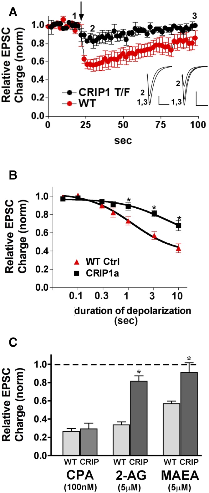
Overexpression of CRIP1a diminishes CB1-mediated DSE in autaptic hippocampal neurons. (A) DSE time-courses for wild-type (WT; red) versus CRIP1a transfected neurons (black) in response to 3-second depolarization (arrow). Insets show sample EPSCs from control (1), maximal DSE inhibition (2), and recovery (3) for CRIP1a-transfected (left) and WT (right) neurons. (B) “Dose” response for DSE using a range of depolarizations from 50 milliseconds to 10 seconds. The wild-type DSE dose response is shown for comparison. *P < 0.05 Bonferroni posthoc test, two-way ANOVA. (C) Bar graph shows relative EPSC charge (1.0 = baseline, no inhibition) after treatment with three drugs under WT and CRIP1a-transfected conditions: CPA, 100 nM; 2-AG (5 μM); MetAEA (5 μM). *P < 0.05, unpaired t test.

