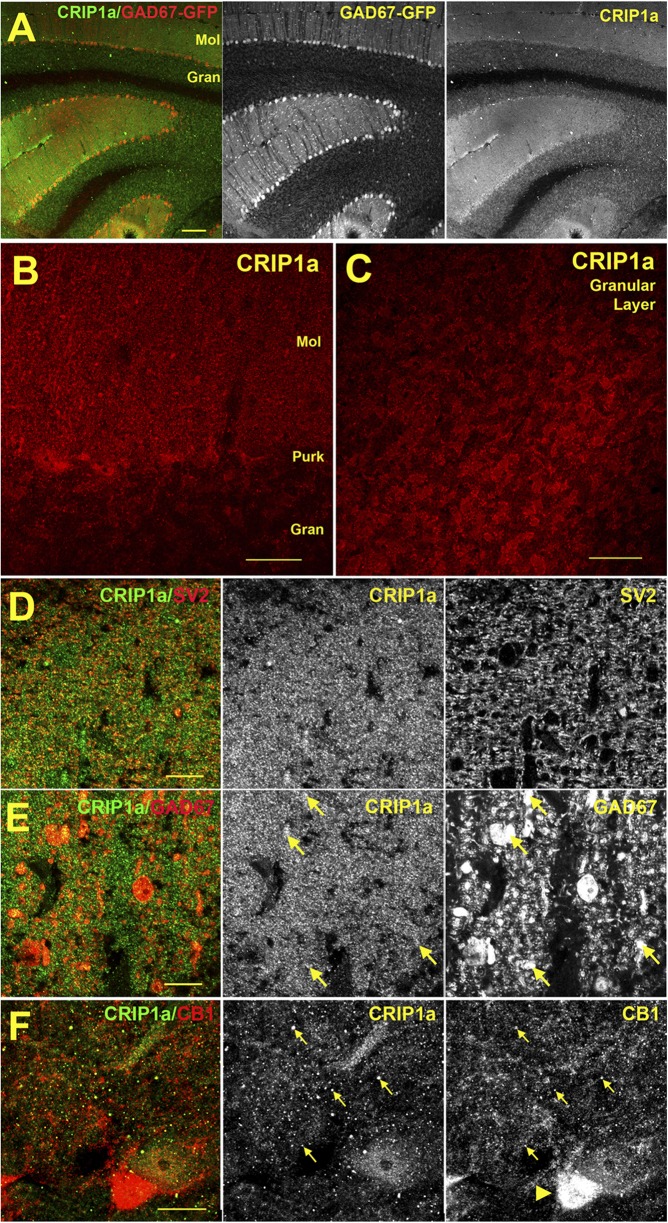
Fig. 2.
Immunohistochemical localization of CRIP1a in the cerebellum of GAD67-GFP transgenic mice. (A) Overview of CRIP1a staining versus GAD67-GFP in murine cerebellum shows a broad distribution in both the molecular (Mol) and granular (Gran) layers. (B) CRIP1a distribution near Purkinje cells. (C) CRIP1a protein in granular layer. (D) CRIP1a overlaps substantially with presynaptic marker SV2 in the molecular layer. In the adjacent image (E), CRIP1a also partially overlaps with GAD67-GFP neurons (arrows), including probable Purkinje cell processes. (F) Sample of CB1R colocalization with CRIP1a (arrows) in the molecular layer near Purkinje cells. Purkinje pinceau region is dense with CB1 (arrowhead). Scale bars, (A) 100 μm; (B) 25 μm; (C) 30 μm; and (D–F) 10 μm.

