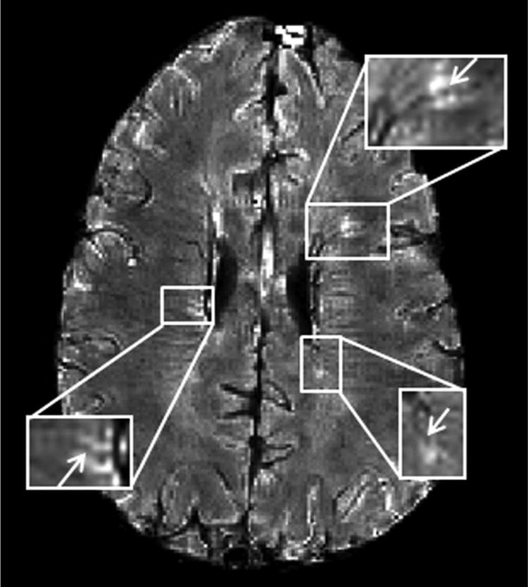
Figure 2.

Co-localization of MS lesions with veins using GEPCI.
T2*-SWI image (1 × 1 × 3 mm3 resolution) in a 33-year-old man with relapsing-remitting MS (EDSS = 4.0, disease duration = 14 years). Three lesions with central veins (arrows) are depicted.
