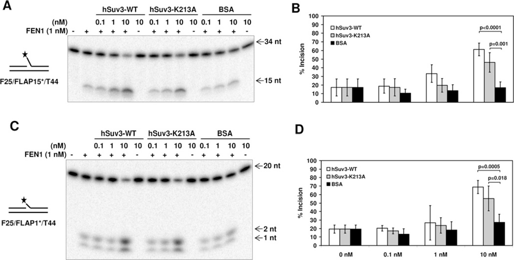
Figure 5. Stimulation of FEN1 incision activity by hSuv3.
(A) FEN1 incision on a long (15 nt) flap in the presence of hSuv3-WT, hSuv3-K213A or BSA. Purified recombinant proteins were mixed (1 nM FEN1) and the reaction started by addition of the radioactively labelled DNA substrate (0.5 nM), followed by incubation at 37°C for 15 min. (B) Quantification of fold stimulation from (A). Error bars represent S.D. (mean value for five experiments). (C) FEN1 incision on a short (1 nt) flap in the presence of hSuv3-WT, hSuv3-K213A or BSA. (D) Quantification of fold stimulation from (C). Error bars represent S.D. (mean value for four experiments). P values were determined using Student’s t test.

