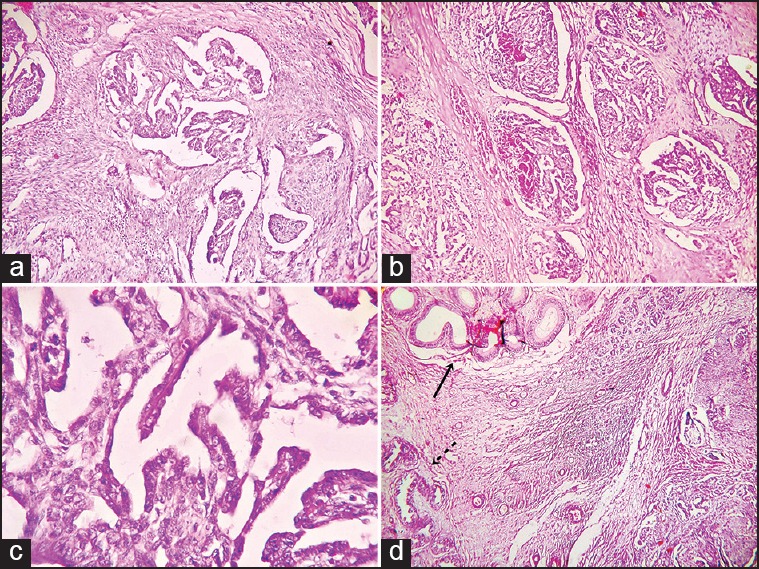
Figure 2.

(a) Biphasic area of the adenocarcinoma showing papillary structures with fibrovascular core along with spindle cell component arranged in fascicles (H and E, ×100) (b) photomicrograph showing epithelial tufts projected into the large dilated epithelium-lined channels of rete testis mimicking renal glomeruli (H and E, ×100) (c) photomicrograph of lining cuboidal to columnar epithelial cells showing moderate pleomorphism (H and E, ×400) (d) photomicrograph demonstrating epididymis (solid arrow) and ductuli efferentes (broken arrow) free from infiltrating tumor mass (H and E, ×100)
