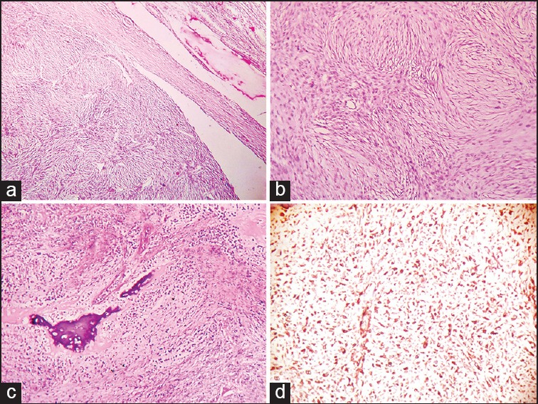
Figure 3.

(a) Photomicrograph of sarcoma-like area of the tumor mass displaying spindle cells arranged in storiform pattern and compressed seminiferous tubules (H and E, ×100) (b) photomicrograph showing spindle cells in storiform pattern, brisk mitotic activity and moderate pleomorphism (H and E, ×400) (c) photomicrograph showing metaplastic bone formation and necrosis in sarcoma-like area (H and E, ×100) (d) photomicrograph showing diffuse and strong positivity of vimentin in sarcoma-like area (×100)
