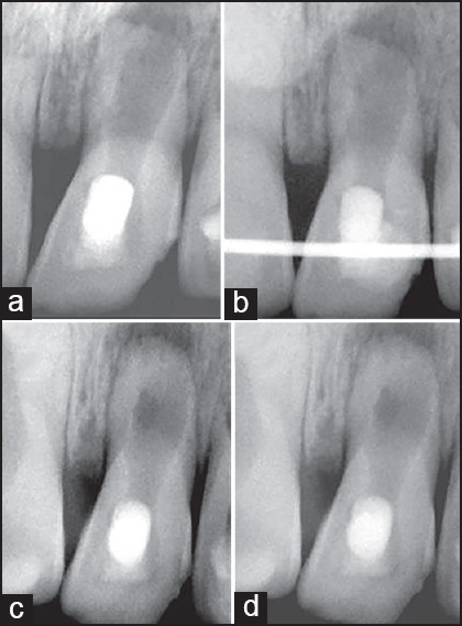
Figure 1.

(a) Tooth #21 exhibited blunderbuss root apex with very thin radicular dentinal walls and diffuse periapical radiolucency. (b) It was stabilised using rigid splinting and PRF aided revascularization was carried out. (c), (d). 12 month and 18 month follow up radiographs revealed excellent periapical healing, apical closure, root lengthening and dentinal wall thickening.
